Overview
BodyMap 3.2, featuring nearly 4,000 anatomical models, introduces several significant enhancements that help students and educators explore the human body more effectively.
In the past 6 months, we added 2D images, 3D animations, and hundreds more 3D body structures to Jack, BodyMap’s full-body virtual avatar, to illustrate the newly discovered muscle layer. We’ve also added an authoritative citation[1] to support these updates.
We will go through each of the exciting, new features and improvements below.
3D Models: 400+ Anatomy Structures Added to Virtual Human Model
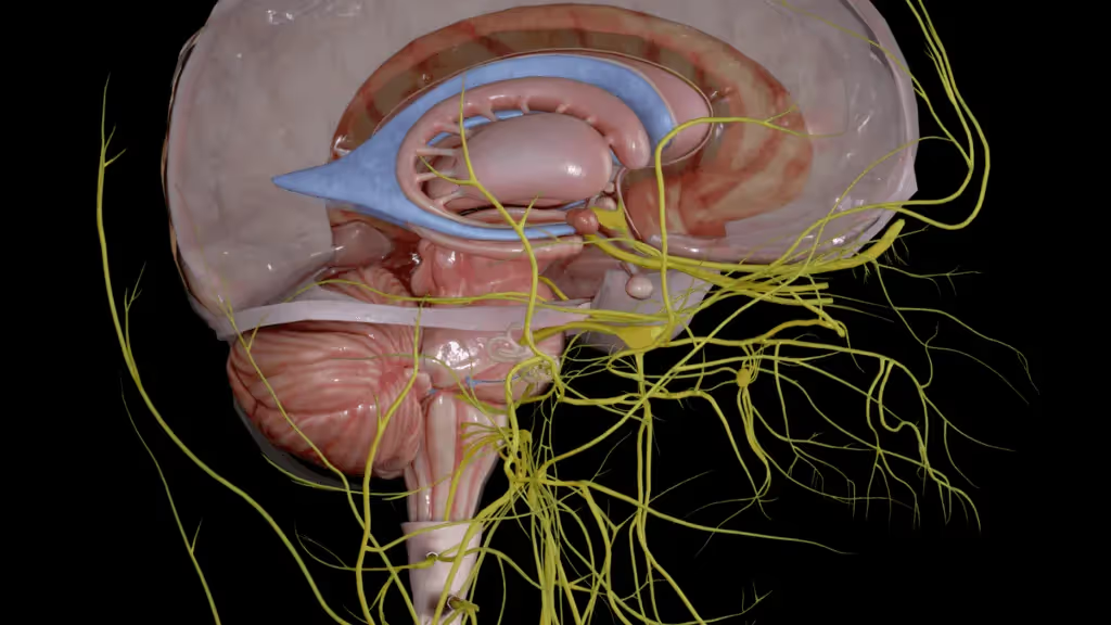
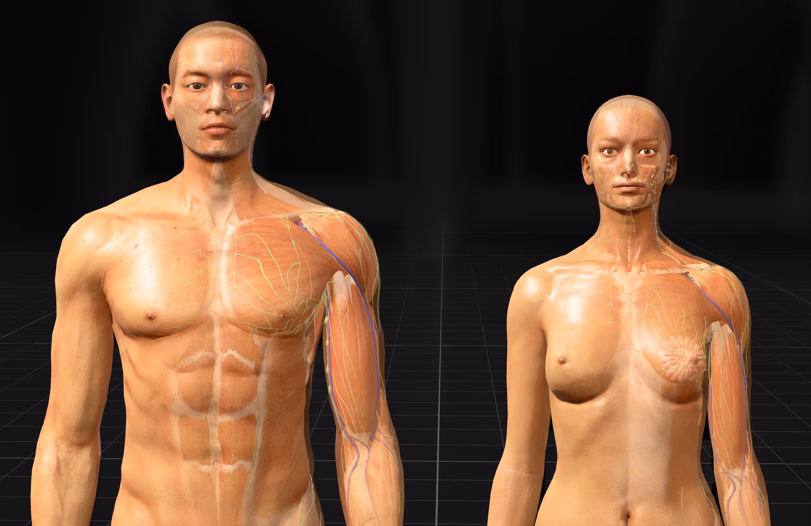
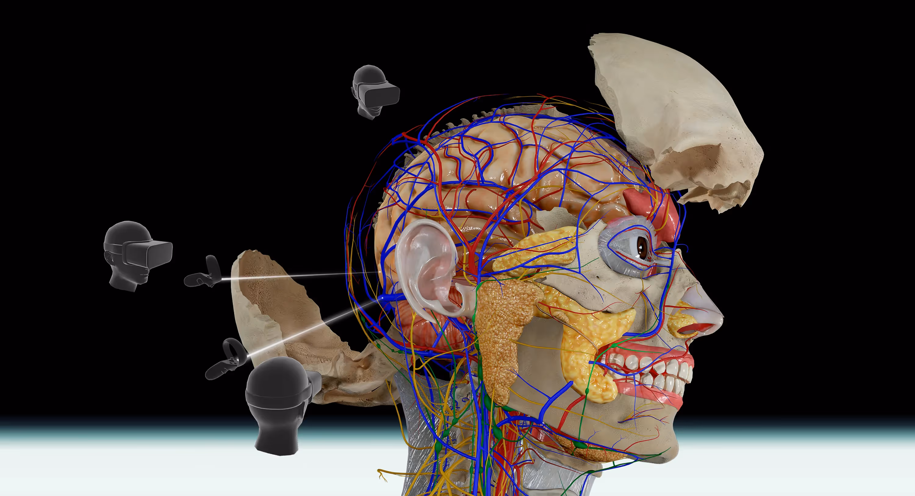
We have added 400+ anatomical structures, including nerves, blood vessels, and muscles, to seven body systems in Jack, BodyMap’s full-body virtual avatar. We also standardized 3D references, aligned muscle naming conventions, and added internal links to connective tissue resources.
For example, you can now observe the esophagus's muscle fibers. You can also view the taste buds on the tongue’s surface.
Another example is the teeth in the skeletal system. Each tooth and its connected nerves are now shown in more detail within the maxillas and mandible.

New 3D models of Esophagus, tongue and more
The fat pads in connective tissues have also been enhanced. The adipose tissue in the cheek region is now more accurately represented, helping students better understand soft tissue structures.
You can zoom in on any body part of our virtual model. Observe clearer and more detailed views at close range.
- Source: MAI press release on BodyMap 3.2 launch, 2024.
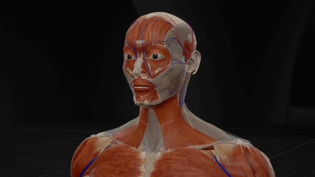
New Muscle Layer Discovered in the Human Jaw
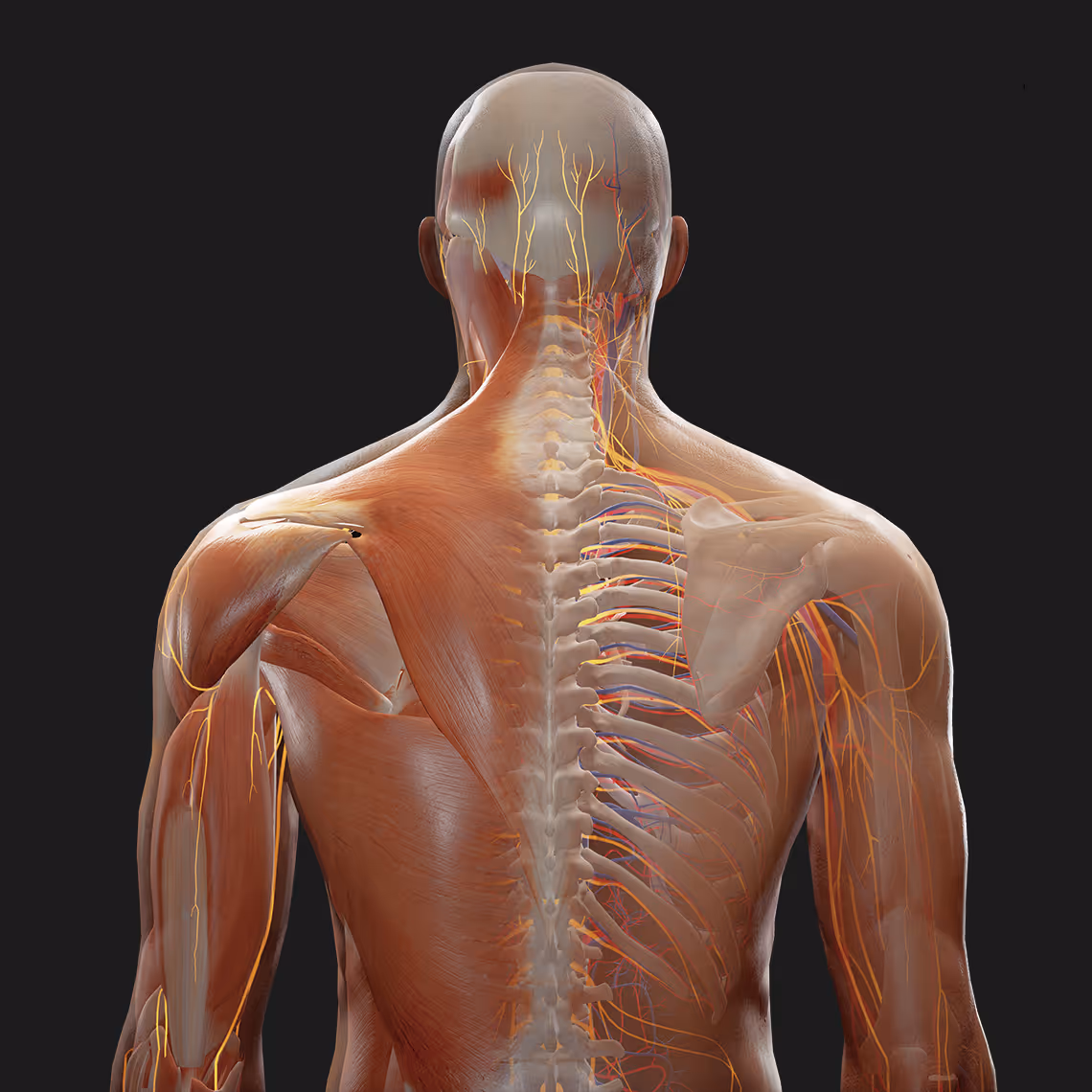
Among the 400+ 3D models added, BodyMap now includes the recently discovered masseter muscle, offering a current, in-depth view for students and educators seeking to stay updated on recent anatomical findings.
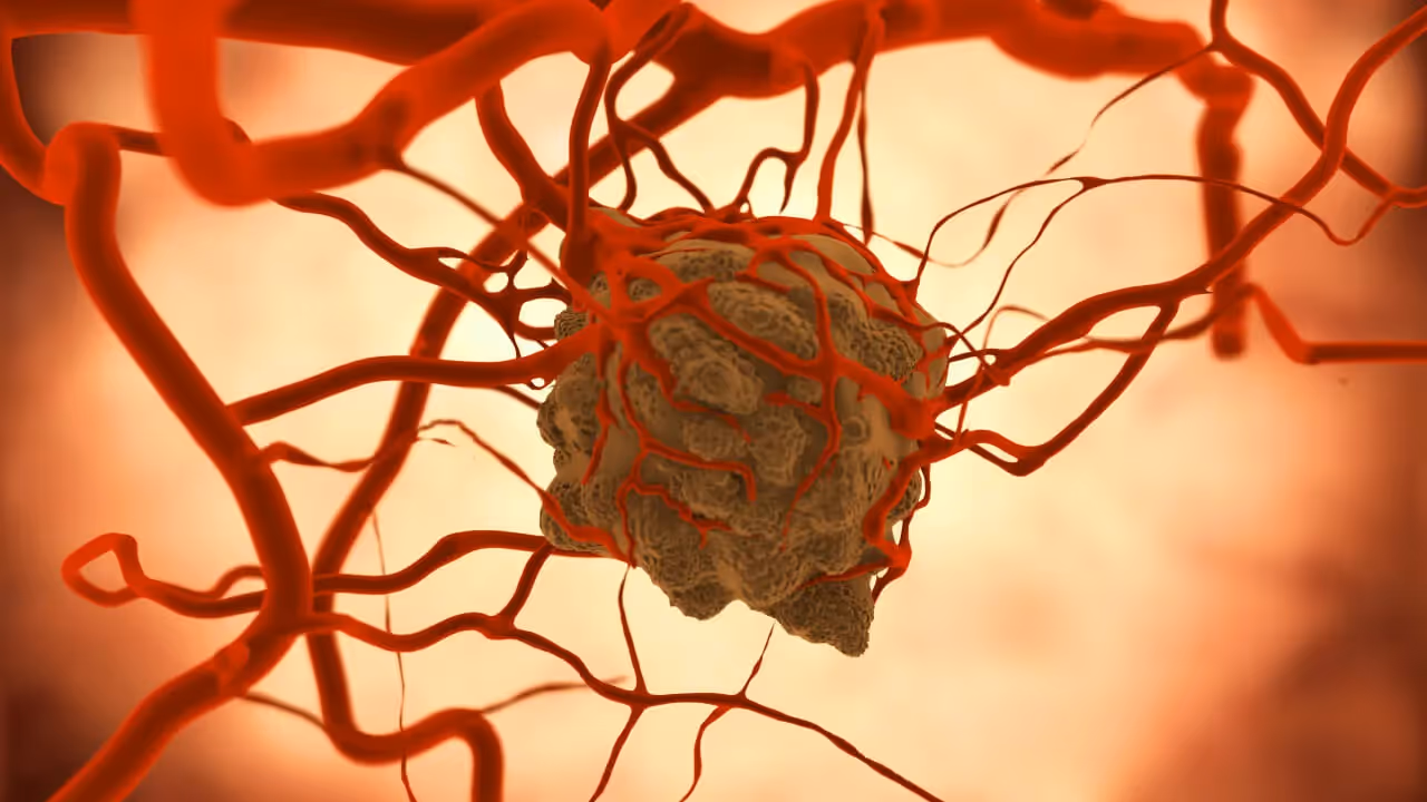
In a research paper published In February 2022, scientists have identified a new layer of muscles at the back of the cheeks in the lower jaw: Musculus masseter pars coronidea (coronoid part of the masseter muscle).
This masseter could be responsible for pulling the lower jaw backward toward the ear, as the researchers pointed out.
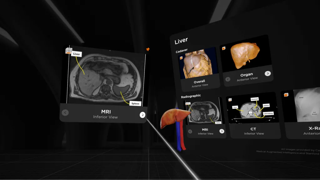
Image Credit: Stanford Medicine Lane Medical Library & NIH National Cancer Institute CIP
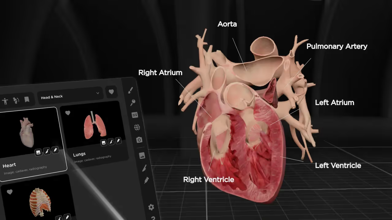
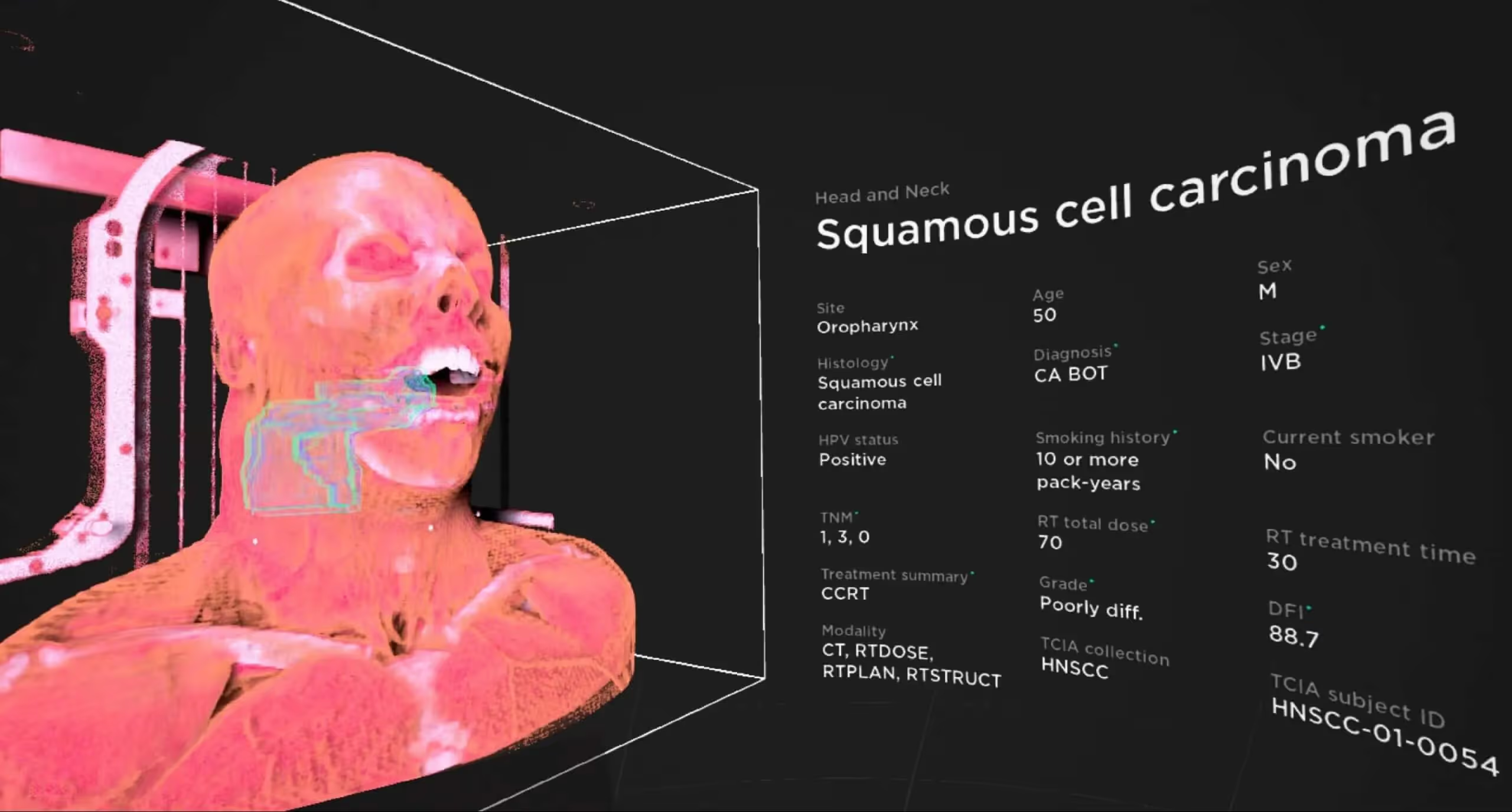
2D Images: Compare Real-Life Scans to 3D Models
BodyMap 3.2 now includes 19 medical images for three organs: Heart, Liver, and Lung. These images are found under the Library tab in the Main Menu. They include:
- Cadaver images
- Radiographic images
Cadaver images show what each organ looks like during dissection. You can also see the organs without surrounding tissues, nerves, or muscles.
The radiographic images include those made using CT (computed tomography), MRI, X-ray, or ultrasound. These can be compared side-by-side with 3D anatomical structures. For certain images, a tooltip defines DICOM, making it easier to learn technical terms.
Comparing 3D anatomical models with real-life 2D images enhances spatial understanding and supports deeper exploration, though learning outcomes may vary. This balanced perspective helps students and educators assess the benefits of each approach.
The 2D images illustrated in BodyMap are from reliable, and scientifically accredited sources such as Stanford Medicine Lane Medical Library and NIH National Cancer Institute CIP (Cancer Imaging Program).
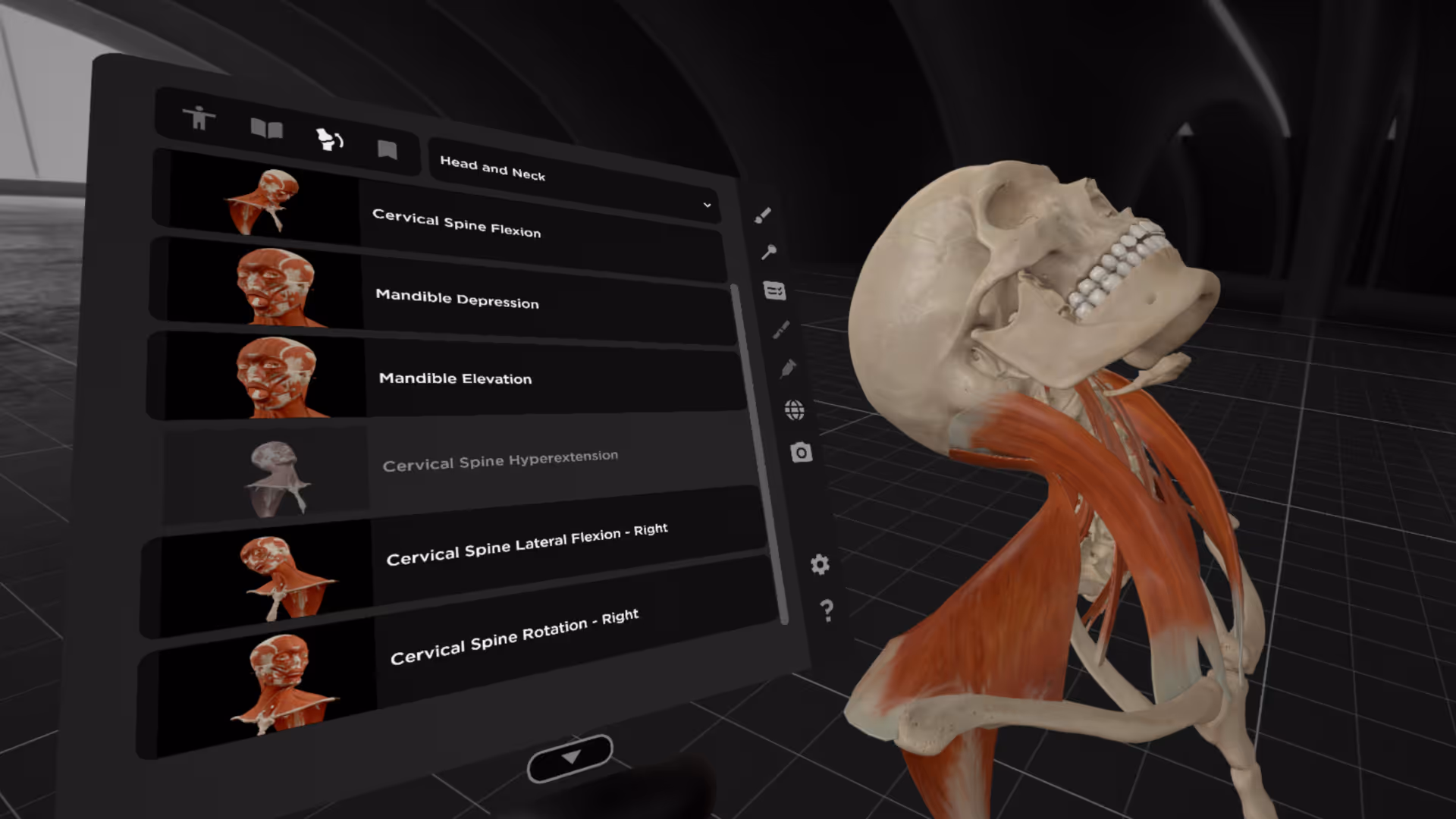
3D Animations: Explore Joint Movements and Muscle Involvement
You will also find a total of 21 movements after you select the Animations tab in your left hand’s Main Menu:
- 14 movements in the right lower limb
- 7 movements in head and neck
Take the neck for instance. When it bends backward, its flashcard will show you the joints and muscles involved in the motion and how much they can move or stretch (range of motion, “ROM”).
Five New Languages Supported in Main Menu
BodyMap’s Main Menu now supports the following languages in addition to English, Simplified Chinese, Traditional Chinese, Japanese and Vietnamese:
- French
- German
- Korean
- Spanish
- Thai
There will be more localized contents in BodyMap in a future release.
Summary
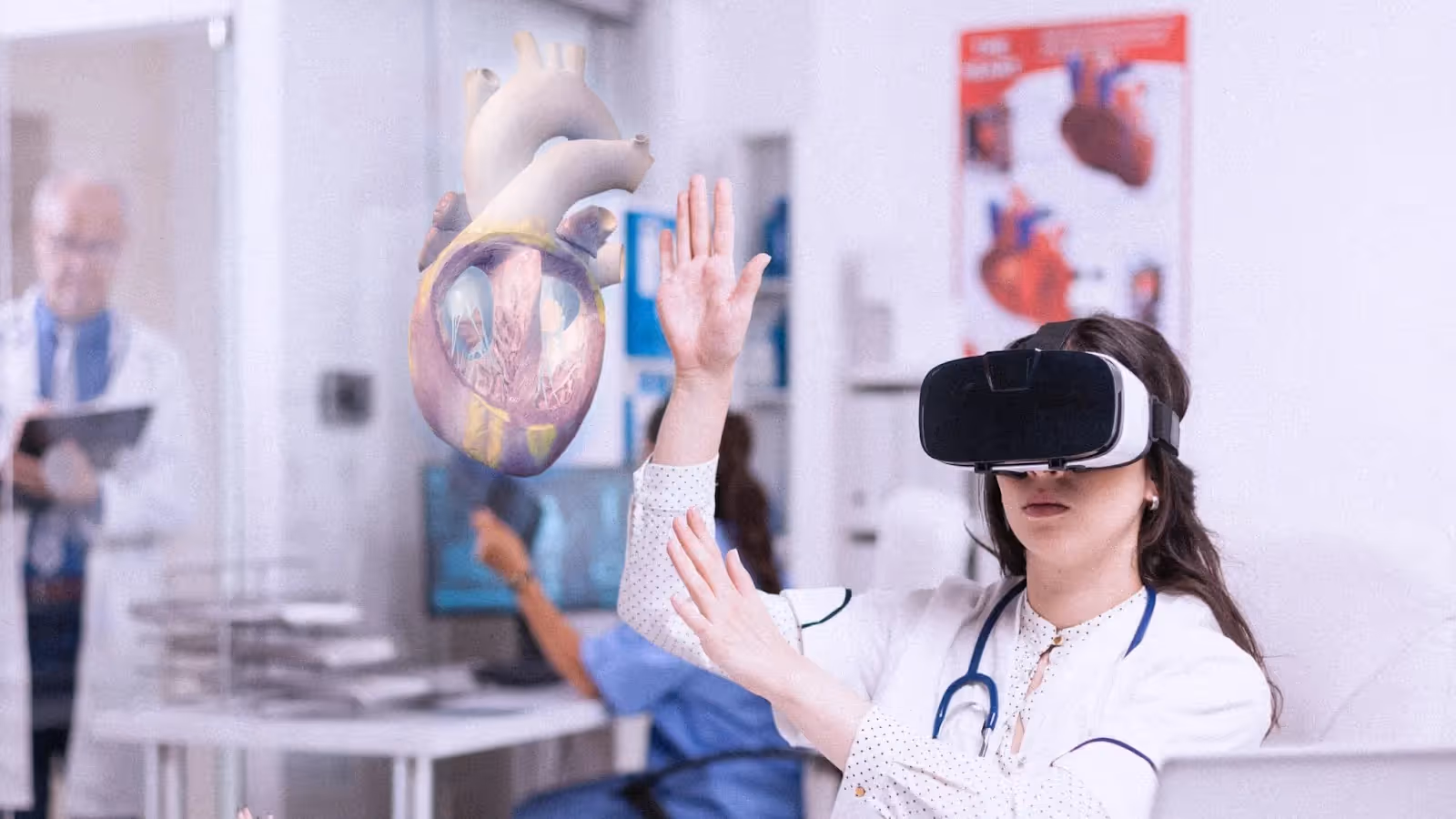
With the upgraded version 3.2, BodyMap currently has over 3,500 anatomical structures in VR, each with high 3D imaging quality and precise information, 19 2D images, and 21 3D animations.
Medical students can now view 2D radiological images (CT, MRI, and X-ray) alongside 3D models, while also observing how joints and muscles move during various motions.
We welcome feedback from educators and students on how BodyMap can better serve your anatomy education needs. Please share your suggestions at hello@mai.ai.
Feedback & Next Steps
We would love to hear what you think! If you have any feedback or suggestions, feel free to reach out. Your insights help us improve BodyMap continually.
Begin your VR anatomy journey today, sign up for a 7-day free trial.
BodyMap
Learn how to navigate the 3D model and utilize the tools to master human anatomy—all in one place.
.jpeg)
.avif)

.avif)
.avif)

The Benefits of Acupuncture for Pain Relief

The Importance of Regular Exercise for Mental Health
The Benefits of Meditation for Stress Reduction
The Importance of a Balanced Diet for Overall Health
The Benefits of Yoga for Mind and Body
The Benefits of Meditation for Stress Reduction