Flat diagrams can only take you so far when the structure you're studying exists in three dimensions. Animated human body models solve this problem by letting you rotate, layer, and explore anatomy the way it actually exists—in space, with depth, and in motion.
This guide covers what animated anatomy models are, how they compare to cadavers and textbooks, and what features to look for whether you're a student, educator, or practitioner choosing a platform for the first time.
What Is an Animated Human Body Model
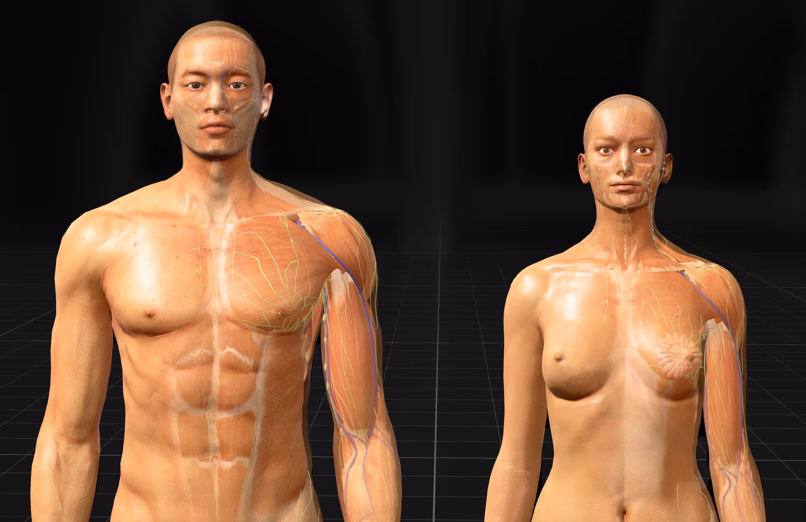
An animated human body model is a digital 3D representation of human anatomy that you can rotate, explore, and interact with in real time. Unlike the static images you find in textbooks, animated models let you peel away layers of tissue, isolate specific body systems, and watch physiological processes like blood flow or breathing happen right in front of you.
What makes a model "animated" is its ability to show movement and change. You might watch the heart contract through its cardiac cycle, observe how muscles shorten during joint flexion, or see the diaphragm move as the lungs expand. This dynamic quality turns anatomy from a memorization task into something you can actually see and explore.
- Animated model: A digital body that can show movement, like circulation or muscle contraction
- 3D anatomy model: A spatial representation you can rotate and view from any angle
- Interactive human body: A tool where you control what you see and how you see it
Animated human body models are now used across medical schools, nursing programs, physical therapy training, and even patient consultations. They offer something flat diagrams simply cannot: a true sense of how structures occupy space and connect to their neighbors.
Why Medical Students and Educators Use Interactive 3D Human Anatomy
The shift toward interactive 3D human anatomy reflects how people actually learn complex spatial information. Textbooks and lectures work well for many subjects, but anatomy involves understanding objects in three-dimensional space—and that's hard to grasp from flat images alone.
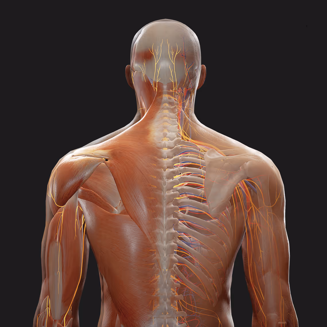
Visualizing Spatial Relationships Between Body Structures
The human body is not a collection of isolated parts. Nerves thread between muscles, blood vessels wrap around organs, and structures layer on top of one another in precise arrangements. Knowing where one structure sits relative to another is fundamental to clinical work.
A 2D cross-section can show you that the brachial artery runs near the median nerve. However, it cannot convey how they travel together down the arm, how their positions shift at different levels, or what the relationship looks like when you rotate your view. A 3D human anatomy model lets you trace these connections continuously, building the kind of mental map that surgeons and clinicians rely on every day.
Think of it like using Google Maps in a new city. You check the street names, notice landmarks, and build a picture in your head of how everything connects. Spatial understanding in anatomy works the same way—you learn where structures are and how they relate to what's around them.
Improving Knowledge Retention Through Active Learning
There's a real difference between reading about anatomy and manipulating it yourself. When you actively rotate a structure, isolate a system, or trace a nerve back to its origin, you engage with the material in ways that passive reading cannot match.
The act of exploration—deciding what to look at, from which angle, at what depth—creates stronger memory traces than following a predetermined sequence of images. You're not just receiving information; you're discovering it.
Reducing Dependency on Cadaver Labs and Physical Models
Cadaver dissection remains valuable, yet it comes with real constraints. Access is limited, specimens cannot be reset once dissected, preservation affects tissue quality, and scheduling dozens of students around a finite number of tables creates logistical headaches.
Digital 3D human body models offer unlimited, repeatable practice without these limitations. You can dissect the same region multiple times, compare normal anatomy to variations, and revisit structures whenever questions come up. This doesn't replace cadaver experience entirely, though it does extend learning opportunities far beyond what physical resources alone can provide.
Features of 3D Human Anatomy Models
Not all anatomy platforms offer the same capabilities. Here's what to look for when choosing a tool for your learning goals.
Interactive Layering and Virtual Dissection
The ability to peel away layers—skin, then fascia, then muscle, then deeper structures—simulates the dissection experience without consuming physical resources. You control the depth, revealing exactly what you want to study while hiding everything else.
360-Degree Rotation and Zoom Controls
Viewing a structure from a single angle rarely tells the whole story. Rotation lets you examine how the back surface differs from the front, how relationships change when you look from above versus below, and how fine details like small openings or muscle attachments appear from different viewpoints.
System Isolation and Structure Highlighting
When you're studying the nervous system, you don't necessarily want every muscle and blood vessel competing for your attention. System isolation lets you focus on one anatomical system at a time, reducing visual clutter and clarifying relationships within that system.
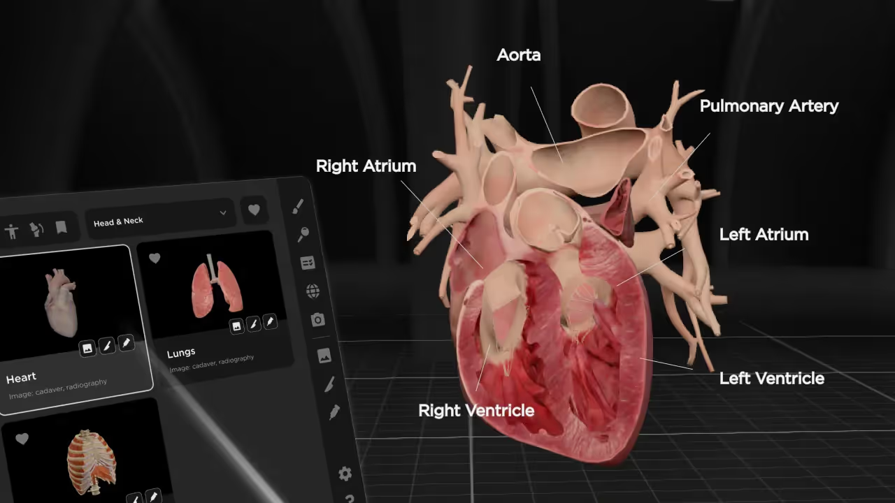
Anatomical Labels and Annotations
Interactive labels do more than name structures. Clicking on a structure might reveal its function, clinical significance, or relationships to neighboring anatomy. This transforms the model from a visual reference into an integrated learning resource.
Animated Physiological Processes
Static models show you what anatomy looks like. Animated models show you what anatomy does. Watching the cardiac cycle unfold, observing peristalsis move through the digestive tract, or seeing respiratory mechanics in action connects structure to function in ways that still images cannot achieve.
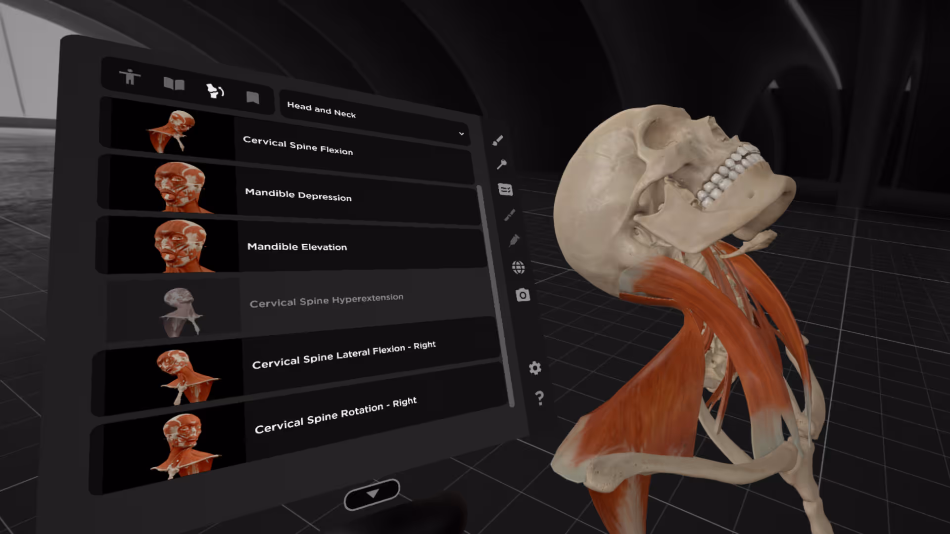
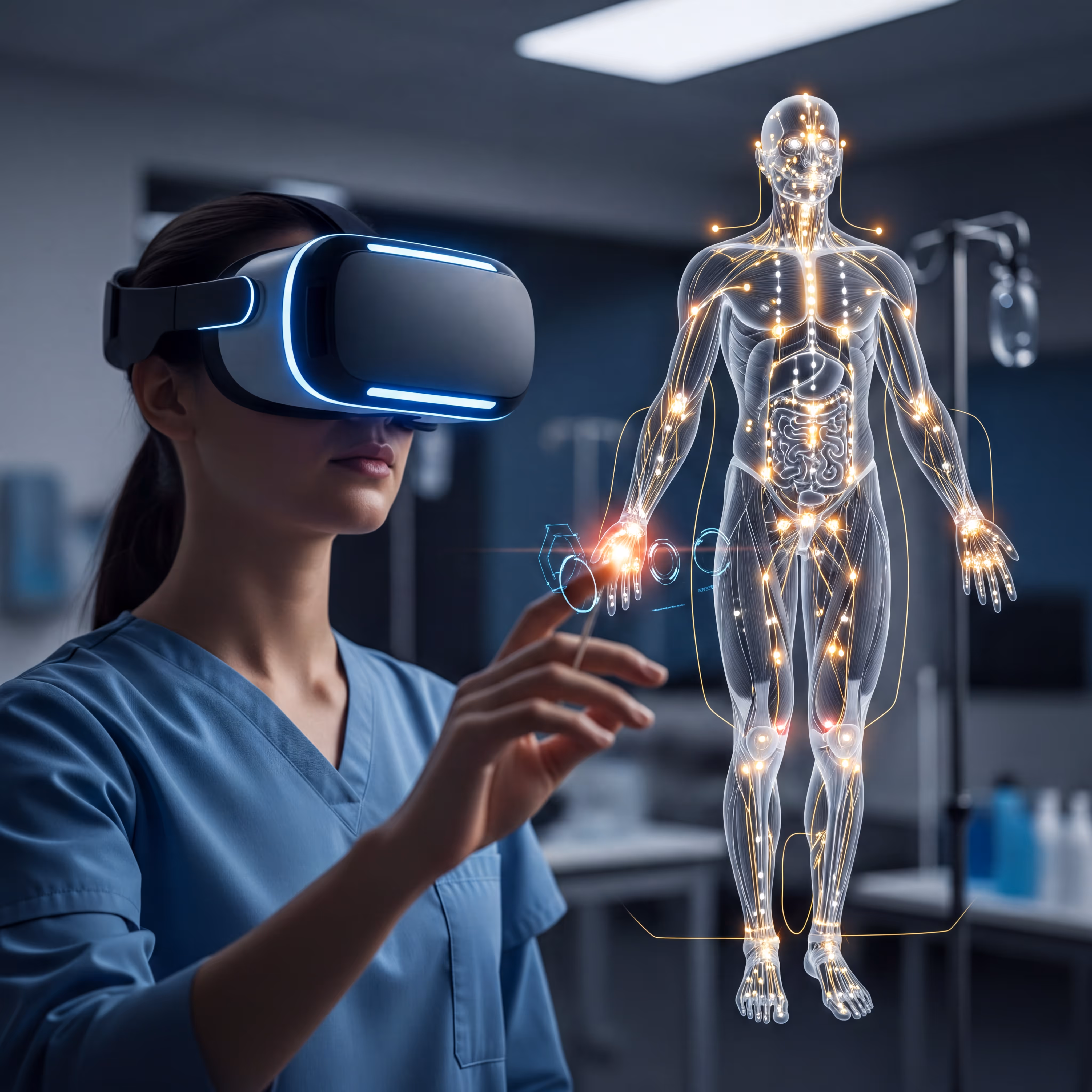
VR and Mixed Reality for Immersive 3D Anatomy Learning
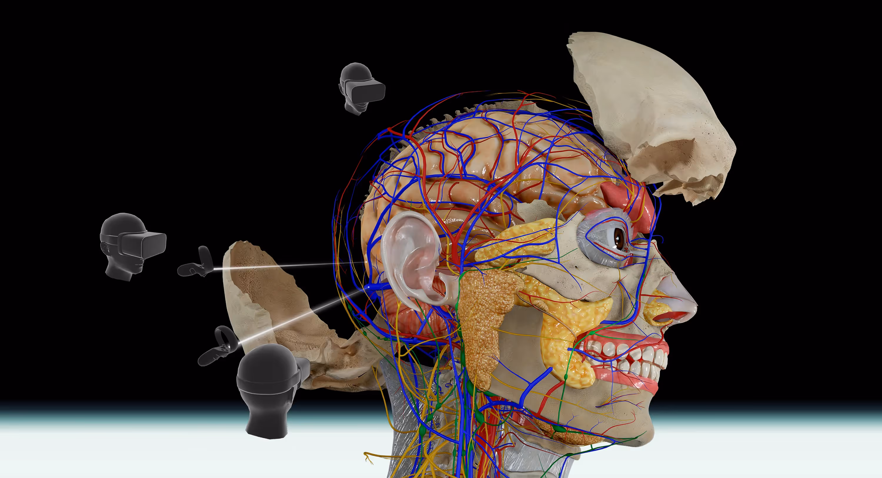
Virtual reality (VR) places you inside a fully digital environment, while mixed reality (MR) overlays digital content onto your physical surroundings. Both technologies take 3D anatomy learning beyond the flat screen and into three-dimensional space.
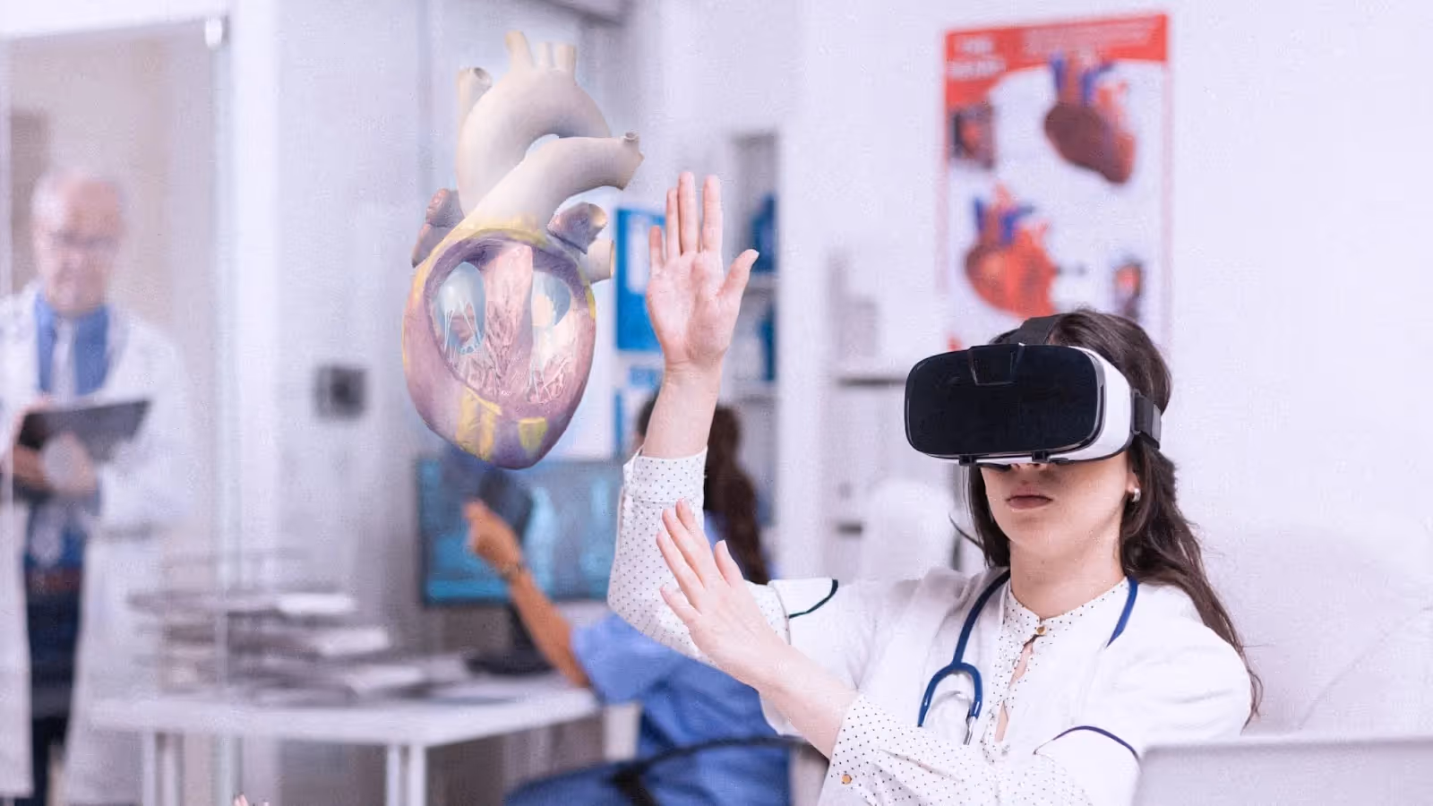
Full Spatial Immersion with VR Headsets
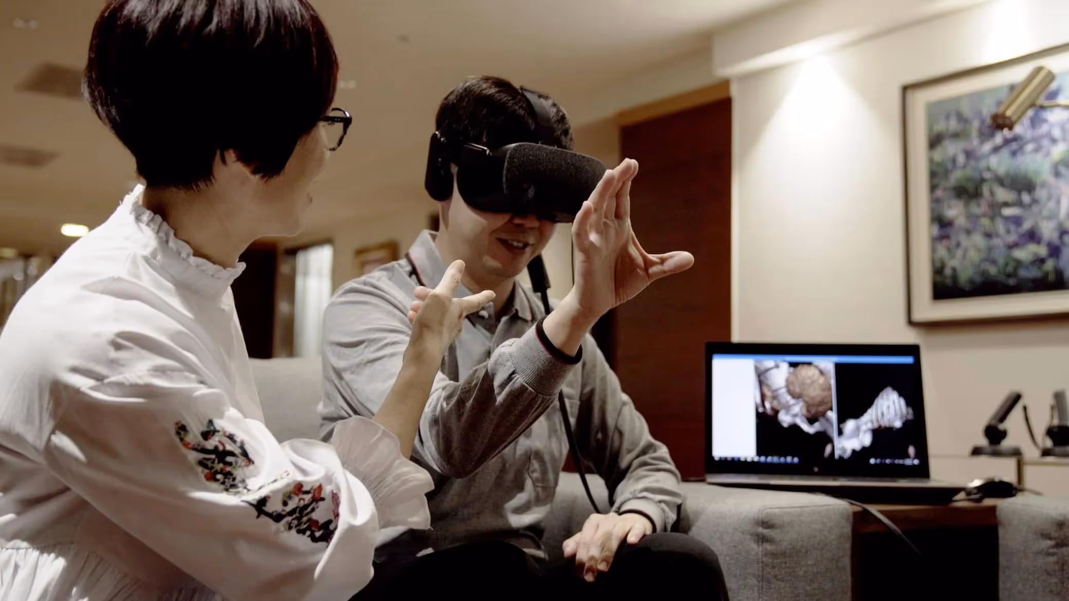
When you put on a VR headset and step into an anatomy environment, structures appear at life size. You can walk around a heart, lean in to examine valve leaflets, or step back to see how the great vessels connect. This spatial immersion builds intuitive understanding that transfers to real clinical encounters.
Platforms like MAI's BodyMap support popular VR headsets including Meta Quest and HTC Vive, making immersive anatomy accessible without specialized lab equipment.
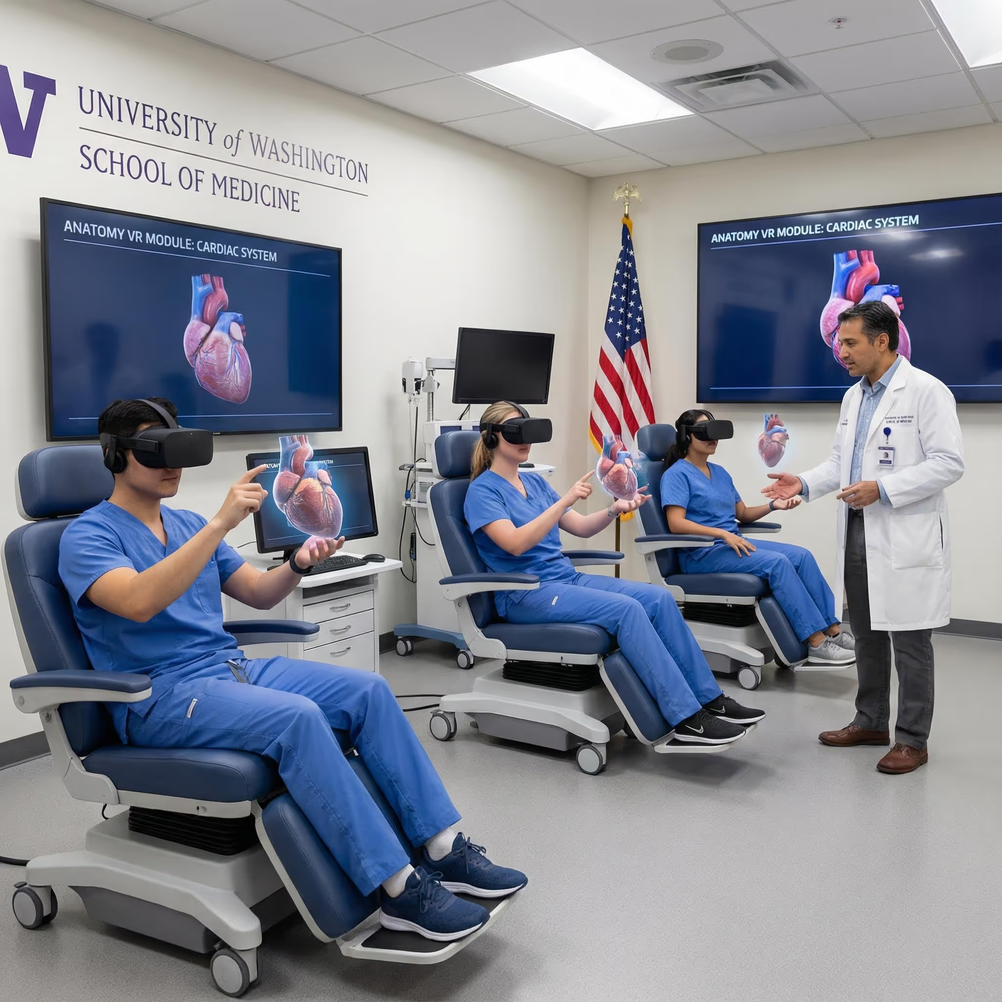
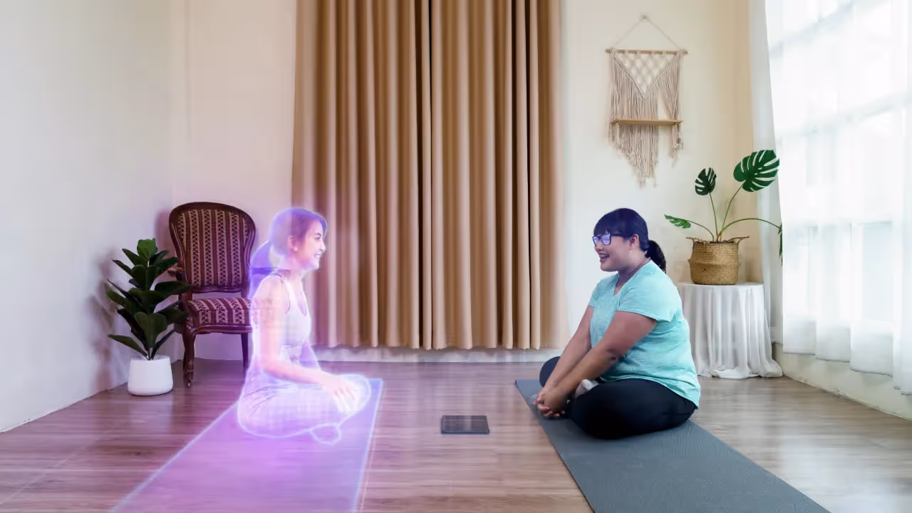
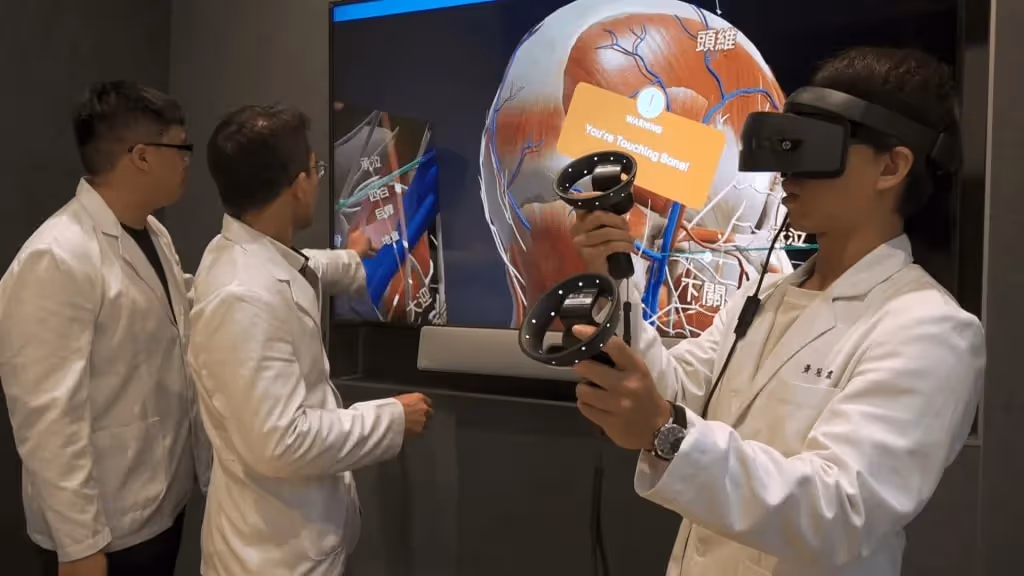
Collaborative Learning with Mixed Reality
MR enables multiple learners to view and discuss the same 3D body model simultaneously in a shared space. An instructor can point to a structure and every student sees exactly what's being referenced—no crowding around a single screen or cadaver table.
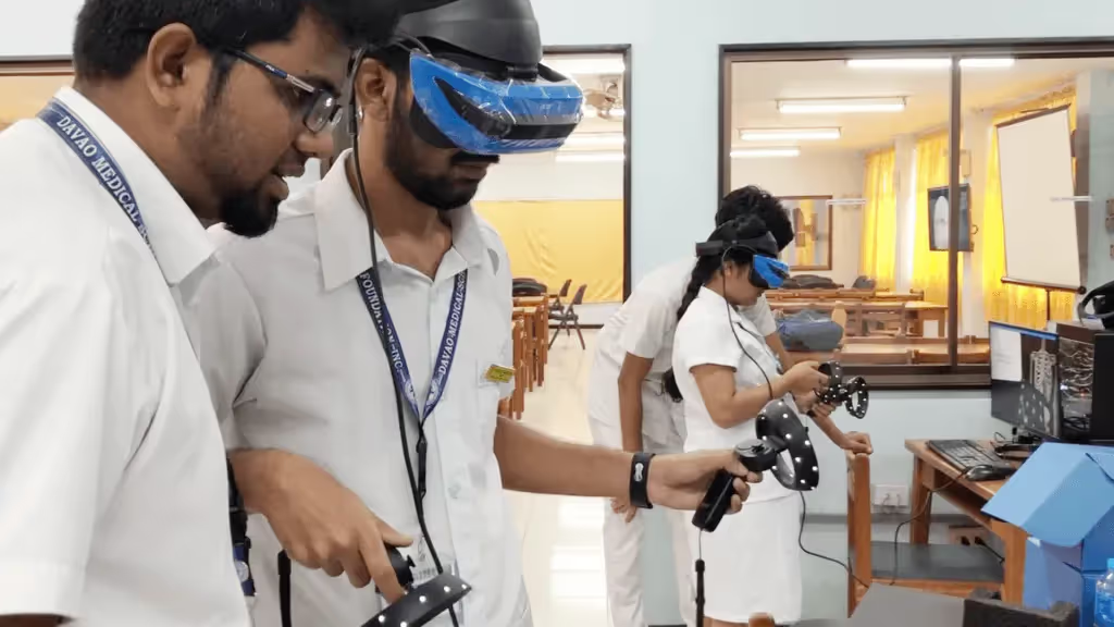
Research on VR Anatomy Learning Outcomes
Studies examining VR anatomy education have found that students report higher confidence and engagement compared to traditional methods. The immersive quality seems to help learners build spatial mental models more quickly, though VR works best as a complement to other learning approaches rather than a complete replacement.
Anatomy Systems in Interactive Human Body Models
Comprehensive platforms cover all major anatomical systems, allowing you to study the body as an integrated whole or focus on specific regions.
- Skeletal system: Bones, joints, cartilage, and their articulations
- Muscular system: Skeletal muscles, tendons, and attachment points
- Cardiovascular system: Heart chambers, arteries, veins, and capillary networks
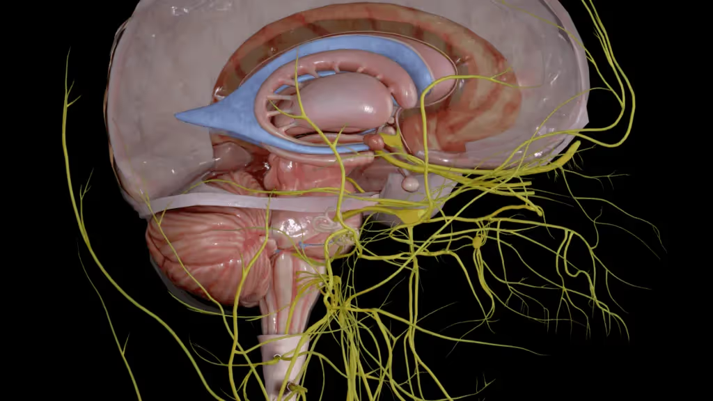
- Nervous system: Brain regions, spinal cord segments, and peripheral nerve distributions
- Respiratory system: Airways, lung lobes, and respiratory muscles
- Digestive system: Alimentary canal from mouth to anus, plus accessory organs like the liver and pancreas
- Lymphatic and immune systems: Lymph nodes, vessels, spleen, and thymus
- Reproductive and urinary systems: Male and female reproductive anatomy, kidneys, ureters, and bladder
- Integumentary system: Skin layers, hair follicles, and associated glands
How 3D Anatomy Models Compare to Cadavers and Textbooks
Each learning method brings distinct strengths. The question isn't which is best overall, but which combination serves your learning goals.
Cadavers provide irreplaceable tactile experience—the feel of tissue planes, the resistance of fascia, the texture of organs. Yet digital models offer something cadavers cannot: the ability to practice the same dissection repeatedly, to undo mistakes, and to study at any hour without scheduling constraints.
Devices That Support 3D Human Body Anatomy Platforms
Your hardware options range from everyday devices to specialized VR equipment, depending on how immersive you want your experience to be.
Desktop Computers and Web Browsers
Many 3D model anatomy platforms run directly in web browsers or as desktop applications. This approach requires no special equipment beyond a reasonably modern computer with a capable graphics card.
Mobile Devices and Tablets
Touch-based interaction on iOS and Android devices makes anatomy study portable. You can review structures between classes, during commutes, or anywhere a laptop would be inconvenient.
VR Headsets Including Meta Quest and HTC Vive
Full immersion requires a VR headset. Consumer devices like Meta Quest offer standalone functionality without a connected computer, while tethered headsets like HTC Vive provide higher visual fidelity when connected to a capable PC.
Choosing the Right 3D Human Body Model for Your Needs
Your ideal platform depends on how you'll use it and what you're trying to learn.
For Individual Medical and Health Science Students
Look for comprehensive anatomy coverage, intuitive navigation, and self-paced exploration features. If you have access to a VR headset, platforms like BodyMap offer immersive experiences that build spatial understanding more quickly than screen-based alternatives.
For Anatomy Educators and Training Institutions
Classroom integration matters here—the ability to display content to groups, track student progress, and align with curriculum objectives. Multi-user support and administrative features become important at institutional scale.
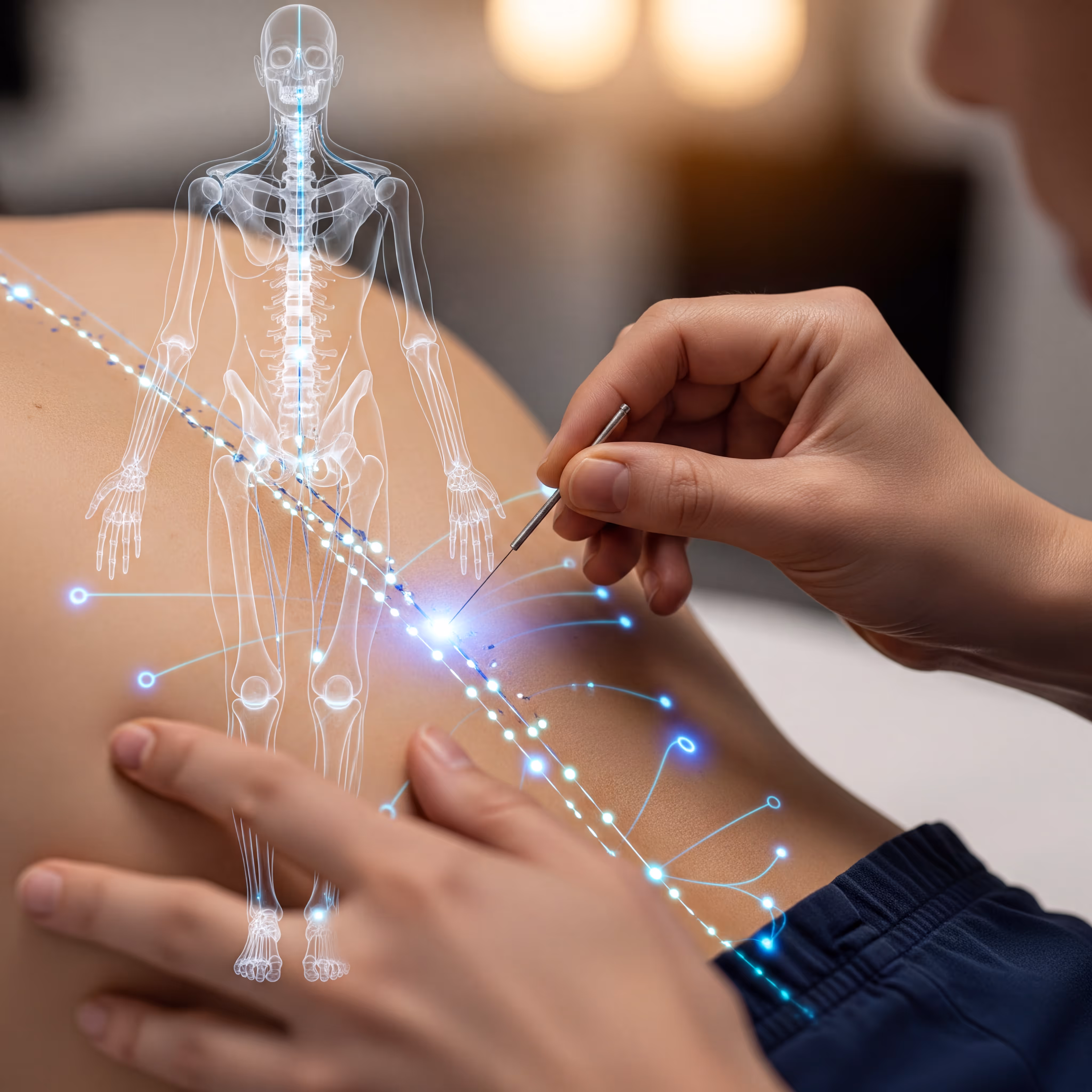
For Acupuncture Schools and Practitioners
Standard anatomy platforms may not include the meridian pathways and acupoint locations that acupuncture education requires. Specialized tools like MAI's AcuMap combine detailed 3D human anatomy with precise acupuncture mapping, serving both foundational anatomy learning and discipline-specific training.
Start Exploring the Human Body with Interactive 3D Anatomy
The shift from static diagrams to interactive, immersive anatomy represents more than a technological upgrade. When you can manipulate structures, trace relationships, and explore from any angle, anatomy transforms from abstract memorization into intuitive understanding.
Start a free trial of the BodyMap anatomy platform
FAQs About Animated Human Body Models
What is the best free 3D human anatomy platform?
Several platforms offer free versions with limited anatomical coverage or restricted features. Evaluate options based on which body systems you need and whether basic labeling meets your depth requirements.
How accurate are 3D human body models compared to real cadavers?
High-quality platforms use anatomically verified data suitable for educational purposes. However, cadavers provide textural and tactile information—tissue consistency, color variation, individual anatomical differences—that digital models cannot replicate.
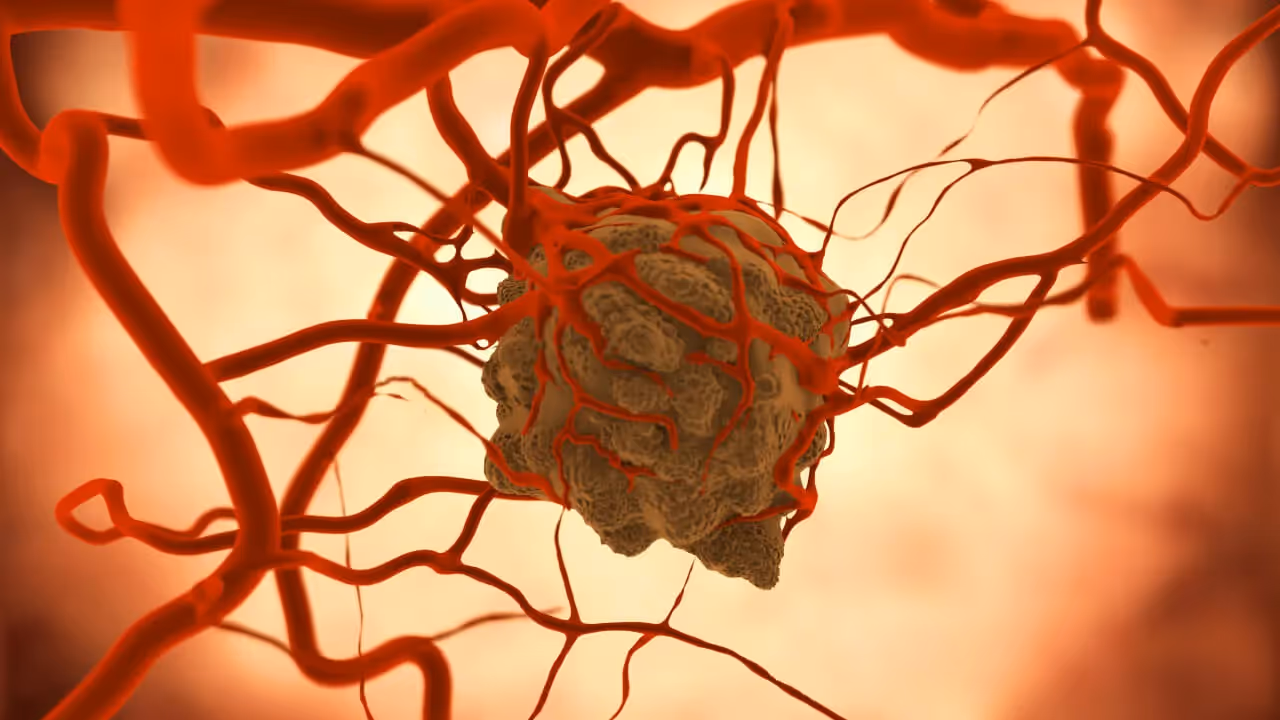
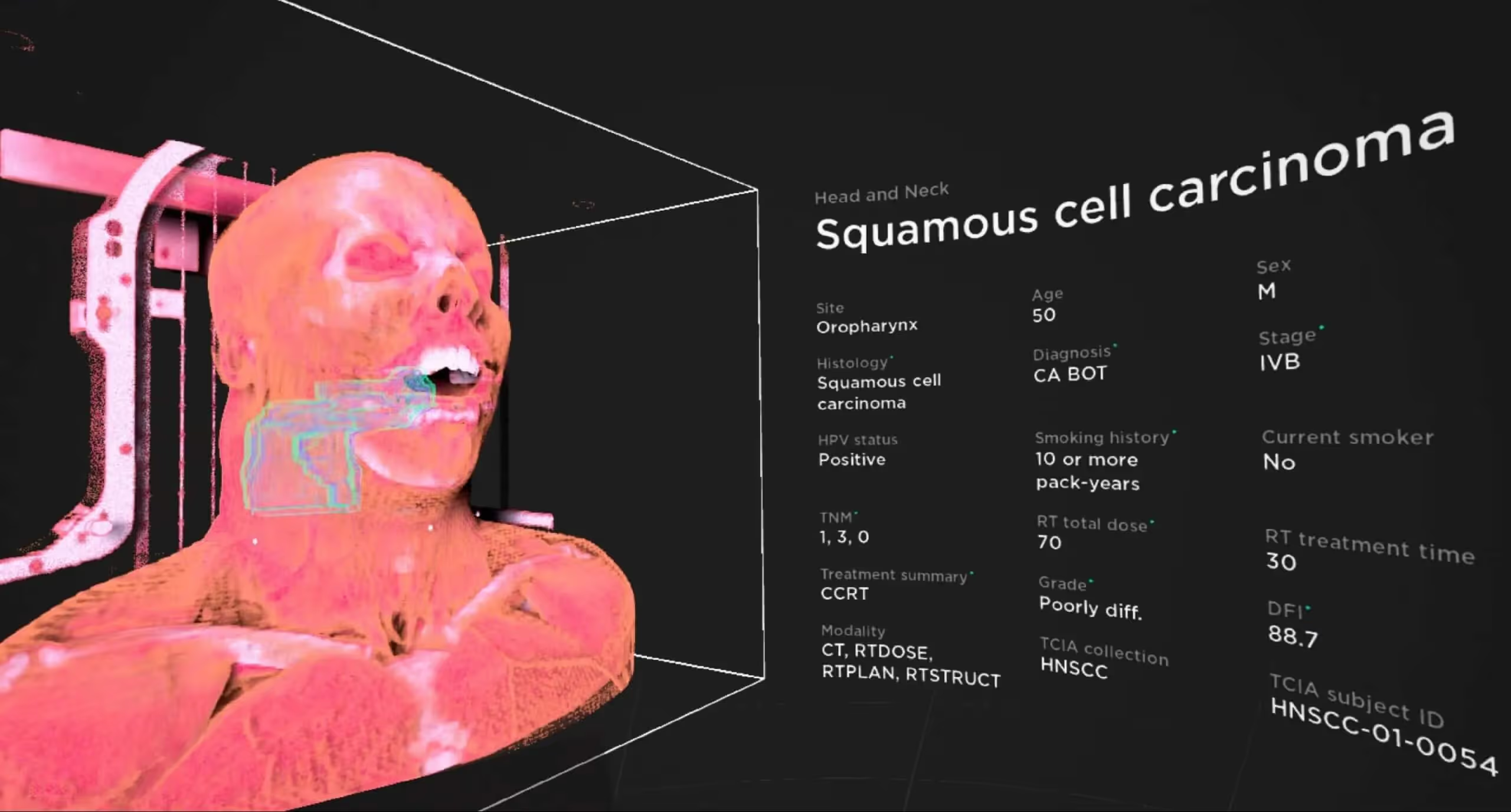
Can animated anatomy models display diseases and pathology?
Some advanced platforms include pathological conditions alongside normal anatomy, showing how diseases alter structure and function. This capability varies significantly between platforms.
What age group benefits most from interactive human anatomy models?
Interactive anatomy tools serve learners from high school biology through professional continuing education. Content depth and complexity vary by platform, with some designed specifically for younger learners and others targeting medical professionals.
Can 3D human body models be used for patient education?
Clinicians increasingly use interactive 3D body models to help patients visualize their conditions, understand proposed procedures, and make informed decisions about treatment options.
Begin your VR anatomy journey today, sign up for a 7-day free trial.
Curriculum Integration Guides
Learn how to navigate the 3D model and utilize the tools to master human anatomy—all in one place.
.jpeg)
.avif)

.avif)
.avif)

The Benefits of Acupuncture for Pain Relief

The Importance of Regular Exercise for Mental Health
The Benefits of Meditation for Stress Reduction
The Importance of a Balanced Diet for Overall Health
The Benefits of Yoga for Mind and Body
The Benefits of Meditation for Stress Reduction