BodyMap 3.2: The visual, and animated VR anatomy learning app
BodyMap 3.2, featuring nearly 4,000 anatomical models, has got a few surprises in store for you.
In the past 6 months, we added 2D images, 3D animations, and hundreds more 3D body structures to Jack, our virtual human model in BodyMap — one of the world’s leading immersive, and interactive medicine VR and healthcare VR solutions for anatomy learning.
We will go through each of the exciting, new features and improvements below.
3D models: 400+ anatomy structures added to virtual human model
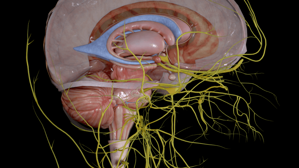
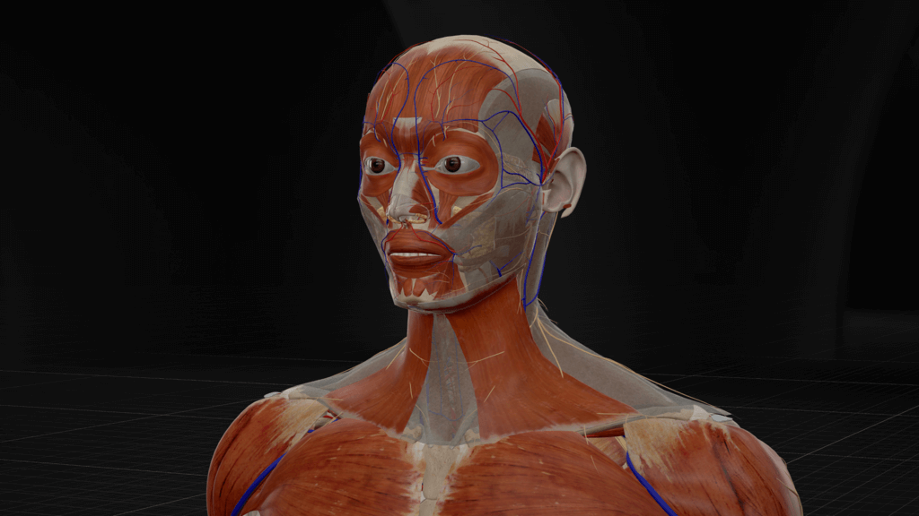
We have added 400+ anatomical structures, including nerves, blood vessels, and muscles, to 7 body systems: digestive, connective, respiratory, muscular, skeletal, nervous, and circulatory systems.
Take for example the esophagus and tongue. You can now closely observe the muscular tube’s vivid muscle fibers, and the taste buds on the tongue’s surfaces.
Another example is teeth in the skeletal system. We now show each tooth and its connected nerves in greater detail in the maxillas and mandible.
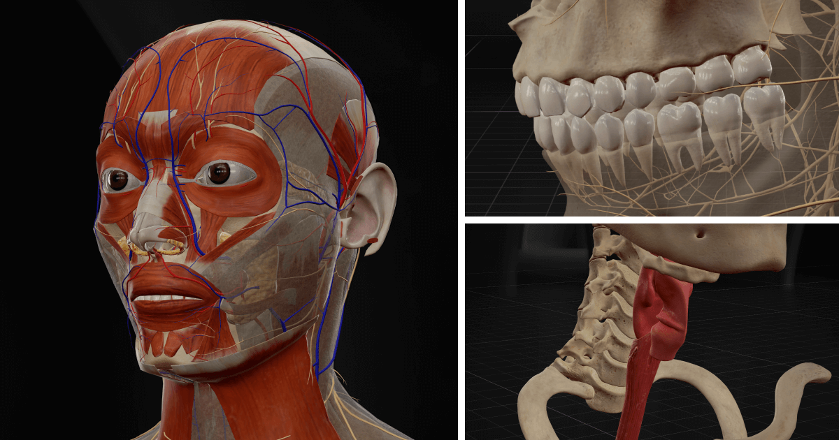
The fat pads in the connective tissues are optimized as well. The yellow mass of fat in the cheeks is now truthful to their real-life counterpart.
You can now further zoom in on any body part of our optimized virtual model, and observe its clearer, and greater details at close-ups.
New addition of a new muscle layer in human jaw
What is worthy to mention here is among the 400+ 3D models added, we are the first to include a newly-discovered masseter.
In a research paper published In February 2022, scientists have identified a new layer of muscles at the back of the cheeks in the lower jaw: Musculus masseter pars coronidea.
This masseter could be responsible for pulling the lower jaw backward toward the ear, as the researchers pointed out.
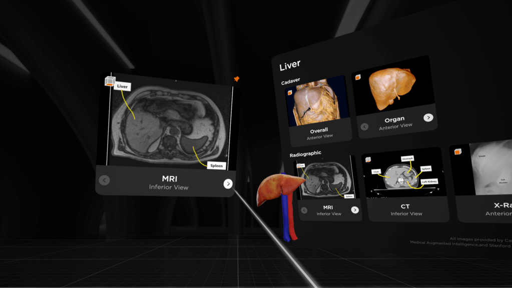
Image Credit: Stanford Medicine Lane Medical Library & NIH National Cancer Institute CIP
2D images: Compare real-life, 2D scans to 3D model
You will find 19 medical images added to 3 organs—Heart, Liver and Lung—including cadaver images and radiographic images, in BodyMap 3.2, after you select the Library tab in the Main Menu.
With cadaver images, you can observe what the organ looks like in a real, dissection session when you cut open the body, and also when it is on its own without its surrounding tissues, nerves, and muscles.
The radiographic images, on the other hand, include those made using CT, MRI, X-ray, or ultrasound. You can compare these 2D, DICOM images to the virtual, 3D model to help speed up your learning process.
You learn more from observing the 3D details of body structures and comparing them to real-life, 2D images than you do from traditional textbooks.
The 2D images illustrated in BodyMap are from reliable, and scientifically accredited sources such as Stanford Medicine Lane Medical Library and NIH National Cancer Institute CIP (Cancer Imaging Program).
3D animations: Watch how joints bend and what muscles are involved
You will also find a total of 21 movements, including 14 movements in right lower limb and 7 movements in head and neck, after you select the Animations tab in your left hand’s Main Menu.
Take the neck for instance. When it bends backward, its flashcard will show you the joints and muscles involved in the motion and how much they can move or stretch (range of motion, “ROM”).
5 new languages supported in Main Menu
BodyMap’s Main Menu now supports French, German, Korean, Spanish and Thai, in addition to English, Simplified Chinese, Traditional Chinese, Japanese and Vietnamese.
There will be more localized contents in BodyMap in a future release.
Summary
With the upgraded version 3.2, BodyMap currently has over 3,500 anatomical structures in VR, each with high 3D imaging quality and precise information, 19 2D images, and 21 3D animations.
Besides radiologists, medical students can also now see body organs’ 2D, radiological images, including CT, MRI and X-ray, in parallel to the 3D model, while observing joints and muscles in motion.
We would love to hear what you think! If you have any feedback or suggestions on where we can go next with BodyMap, let us know at[email protected].